Chronic knee pain is a common complaint among older adults, and it can have many causes, ranging from simple overuse to more complex underlying conditions. When a 65-year-old woman comes into a clinic after months of struggling with knee pain that doesn’t improve despite trying various treatments—including traditional Chinese medicine—it’s time for a deeper look.
In her case, after trying multiple treatments without success, the decision was made to take an X-ray during her visit. Based on this clinical scenario, let’s explore the most likely causes of her knee pain and the potential diagnosis.
A Look at the Key Symptoms
- Chronic Knee Pain: The fact that the pain has persisted for months suggests this is not a simple, acute injury like a sprain or strain.
- Treatment Resistance: The fact that traditional medicine and other therapies didn’t provide relief hints at a more complex or structural issue in the knee.
- Age Factor: At 65, the likelihood of age-related wear-and-tear on the joints increases, as does the potential for degenerative conditions like osteoarthritis.
Common Causes of Chronic Knee Pain in Older Adults
- Osteoarthritis (OA) 🦵
Osteoarthritis is one of the most common causes of knee pain in older adults. It’s a degenerative joint disease where the cartilage protecting the bones in the knee breaks down, leading to pain, stiffness, and sometimes swelling. OA develops gradually over time and is worsened by age, previous injuries, or repetitive stress on the joints.
Symptoms of OA include:
- Pain that worsens with activity (walking, standing, climbing stairs)
- Stiffness, especially after periods of rest
- Swelling or tenderness around the joint
- A feeling of instability in the knee
- Limited range of motion
An X-ray would typically show joint space narrowing, bone spurs (osteophytes), and subchondral sclerosis (increased bone density underneath the cartilage).
- Rheumatoid Arthritis (RA) 🧑⚖️
Rheumatoid arthritis, an autoimmune condition, can also cause chronic knee pain. It occurs when the body’s immune system mistakenly attacks the joints, leading to inflammation, pain, and damage. RA usually affects both knees, but the pain may not be symmetrical in every case. The pain can be especially intense in the morning or after periods of inactivity.
Symptoms of RA include:
- Symmetrical joint pain (both knees affected)
- Morning stiffness that lasts for more than an hour
- Swelling and redness around the joints
- Decreased range of motion
X-rays of RA often show joint erosion, soft tissue swelling, and bone deformities as the condition progresses.
- Patellofemoral Pain Syndrome (PFPS) 🎾
Also known as “runner’s knee,” PFPS occurs when there is pain around the kneecap (patella) due to overuse, misalignment, or improper tracking of the patella as the knee moves. While this condition is more common in younger individuals, it can also affect older adults, especially those who have been relatively inactive but suddenly increase their activity level.
Symptoms include:
- Pain in the front of the knee, around the patella
- Pain that worsens with activities like running, climbing stairs, or squatting
- A feeling that the knee might “give way” during activity
X-rays of PFPS typically show no major abnormalities, but they may reveal patellar malalignment or chondromalacia patellae (softening of the cartilage beneath the patella).
- Meniscus Tears 🩹
A meniscus tear is another possibility. The menisci are cartilage structures in the knee that act as shock absorbers. As we age, these menisci can wear down or tear from an injury or wear-and-tear over time, causing pain, swelling, and difficulty moving the knee. While some meniscus tears are caused by trauma, others can occur gradually, particularly in older adults due to degenerative changes.
Symptoms of a meniscus tear include:
- Pain in the knee, especially along the joint line
- Swelling and stiffness
- A sensation of the knee “locking” or “catching”
- Difficulty fully straightening or bending the knee
X-rays won’t always show a meniscus tear, but an MRI would reveal the tear in detail. However, X-rays can help rule out other conditions like fractures or arthritis.
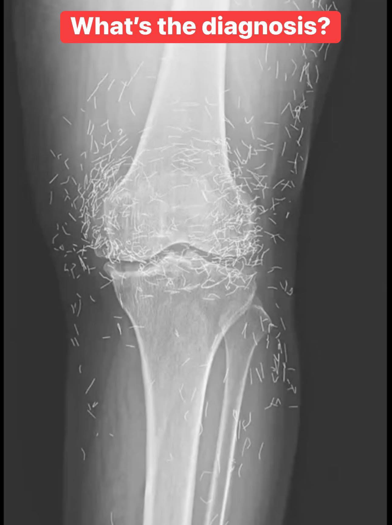
The X-ray – A Key Diagnostic Tool
X-rays are typically the first imaging tool used to evaluate chronic knee pain. They allow doctors to see structural issues in the bones, joint spaces, and alignment of the knee. Based on the findings from the X-ray, we can narrow down potential diagnoses.
Possible X-ray Findings
- Osteoarthritis: The X-ray may reveal joint space narrowing, osteophytes (bone spurs), and subchondral sclerosis. These are signs of cartilage loss and bone changes associated with OA.
- Rheumatoid Arthritis: The X-ray may show joint erosion, soft tissue swelling, and possibly even deformities in the joint alignment.
- Meniscus Tears: While meniscus tears may not be directly visible on an X-ray, other changes, such as bone bruising or joint alignment issues, could be present.
- Patellofemoral Pain Syndrome: This condition often doesn’t show up on an X-ray, but the doctor might look for signs of misalignment, cartilage wear, or soft tissue swelling.
Conclusion: What Might Be the Diagnosis?
Given the patient’s age and the fact that her knee pain has been chronic and resistant to multiple treatments, osteoarthritis (OA) is the most likely diagnosis. The X-ray would likely show signs of cartilage breakdown, joint space narrowing, and possibly osteophytes (bone spurs) in the knee joint.
While other conditions like rheumatoid arthritis, patellofemoral pain syndrome, or meniscus tears could also be possible, OA is the most common cause of chronic knee pain in older adults, especially when it resists treatment.
The good news is that while OA is a degenerative condition, there are various treatment options to help manage symptoms and improve quality of life, including physical therapy, pain management strategies, joint injections, and, in some cases, surgery (like knee replacement).
So, if you or someone you know is experiencing chronic knee pain, don’t hesitate to consult with a healthcare provider. Getting an early diagnosis and starting treatment can help prevent further joint damage and improve mobility over time.
